Clinical picture
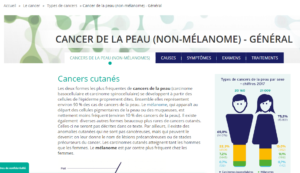
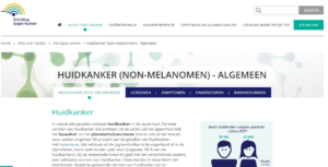
Basal cell carcinoma (BCC) is the most prevalent and least malignant type of skin cancer. BCC tumours originate in the epidermis and can occur all over the body. However, in most cases BCC tumours are found in areas that receive the most sun exposure, such as the face.
Even though BCC tumours are very slow-growing and hardly ever spread, treating the cancer remains important. The tumour can affect surrounding tissues, thus greatly diminishing the outlook.
Dermatologists and oncologists identify several different types of BCC.
- Nodular BCC is the most common type. It represents almost 70% of all cases. Clinically it presents as a well-defined translucent pearly nodule that is either round or oval with a rolled border and occasional ulceration. Most tumours of this kind are observed on the face. The micro-nodular subtype is a cluster of nodular tumours close to one another.
- Superficial BCC is the second most common subtype of BCC (18% of all cases). It presents as slightly elevated plaque that may be scaly, most often developing on the torso or shoulders.
- Pigmented BCC is a rare variant (7% of all cases). It ranges from brown to blue black and can be mistaken for melanoma.
- Morphea-like (or morpheaform) BCC is a rare, aggressive variant accounting for less than 4% of all our BCCs. It presents as firm plaques that are yellow or white with ill-defined borders. The extent of the tumour is usually not apparent on clinical examination.
- Mixed type BCC: a combination of two or more types of BCC.
BCC is the fastest growing cancer type in Belgium. In 2003, 8,000 cases were diagnosed; that number more than tripled to over 25,000 in 2015. BCC makes up around 75% of all skin cancer patients. It is mostly found in patients over 60 years old. The mortality rate is very low.
Symptoms
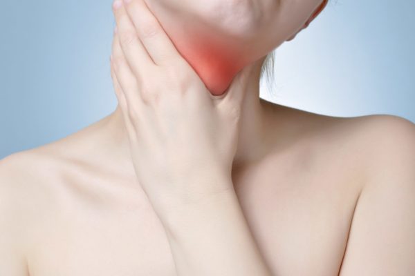
Most of the time, a BCC tumour will manifest itself as a smooth little lump on the skin. Sometimes a small lesion may develop that does not heal very quickly. It will form a scab that will fall off eventually.
Cause
There are several known factors that increase the risk of contracting a basal cell carcinoma. These include:
- Ultraviolet radiation: excessive exposure during childhood and the number of times a patient has had sunburn all contribute to the possibility of developing a BCC tumour later in life.
- Skin type: Fair-skinned people run a greater risk of developing skin cancer.
- Compromised immune system: the negative effect of UV radiation is enhanced when the immune system is diminished.
- Radiation history: if someone has received radiation therapy at a young age, the treated skin area will carry a greater risk of developing skin cancer.
- Light therapy: patients that are being treated with PUVA light therapy have an elevated risk of contracting skin cancer.
- Hereditary factors: Between 1 and 5% of patients has basal cell naevus syndrome (BCNS, Gorlin’s Syndrome). BCC tumours will develop faster in this patient group.
- Medical history: having had any type of skin cancer before.
- Hereditary dermatological conditions: conditions such as xeroderma pigmentosum, epidermodysplasia verruciformis, recessive dystrophic epidermolysis bullosa, albinism and polymorphism all bring an elevated risk of skin cancer.
- Smoking doubles the risk of contracting skin cancer.
Diagnosis
If a GP suspects a patient may have skin cancer, he or she will be referred to a dermatologist. The dermatologist will do further research, such as a biopsy where some tissue from the affected skin area is removed. This will help determine the BCC type with which the patient is dealing. Alternatively, the dermatologist may opt to excise the entire tumour under local anaesthetic.
There is a distinction between low-risk and high-risk BCC tumours. High-risk tumours are micronodular tumours, morphea-like tumours, tumours that are larger than 2 centimetres across, recurring tumours and tumours in the face.
Treatment
Depending on the tumour type, stage and location, the dermatologist will come up with a treatment strategy. Often, this involves operative removal of the tumour, sometimes followed up by radiation sessions. When the tumour cannot be removed completely, subsidiary treatment is necessary. Methods include:
- radiotherapy
- cryosurgery (freezing of the tumour)
- photodynamic therapy
- Mohs-surgery (scraping off tumour layers and laser removal)
- chemotherapy
- immune therapy
Hedgehog pathway inhibitors, for eligible patients.
Additional information
Patient organisations
Related articles
Clinical picture
Symptoms

Cause

Diagnosis

Treatment

Links