Clinical picture
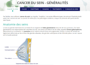
Breast cancer is the collective name for a malignant cell growth in the breast. When normal cells form, spontaneous genetic mutations can take place. Normally, the immune system is able to deal with a compromised cell, but sometimes these cells remain and grow into a tumour. Breast cancer can develop anywhere in the breast and afflicts both men and women, although the disease is far more prevalent in women.
For women, breast cancer is the most frequently occurring type of cancer. In Belgium, 10,500 new cases are diagnosed every year, and every year more than 2,300 women die of breast cancer. It is estimated that one in every nine women will contract the disease before she is 75 years old, and 75% of cancer patients are over 50 years old when diagnosed.
Because breast cancer is relatively treatable when diagnosed in time, Belgium has put a national screening program in place. Every Belgian woman between the ages of 50 and 69 is invited to have a mammogram taken once every two years.
In around 20 to 25% of cases, the cancer spreads beyond the breast area. Once breast cancer has metastasised, it becomes much harder to cure. In most cases, only palliative care is offered in order to make the remaining months or years easier.
There are many different types of breast cancer, depending on where the tumour develops, whether it grows and at what rate and whether treatment is possible. The most common types of breast cancer are:
- Ductal carcinoma in situ (DCIS): This type of cancer develops in the milk tubes of the breast and does not spread. It occurs in around 15% of all cases.
- Invasive ductal carcinoma: This is the most prevalent form of breast cancer, in some cases it can start off as a DCIS tumour. An Invasive ductal carcinoma can, as the name indicates, spread to surrounding tissue.
- Lobular carcinoma in situ (LCIS): This tumour develops within the milk gland and does not spread to surrounding tissue. Patients rarely experience any symptoms, and this cancer type usually gets detected by chance or by the national testing programme.
- Invasive lobular carcinoma: This type can develop from an LCIS tumour, but unlike LCIS this tumour type can grow and spread to other organs and tissues. It occurs in 10% of all cases.
- Hormone-sensitive breast cancer: When a tumour grows under the influence of oestrogen or progesterone, we call it a hormone-sensitive tumour. These tumours carry a lower risk of spreading, and the outlook is relatively good. Around three-quarters of women with breast cancer have a hormone-sensitive tumour.
- HER2-positive breast cancer: This type contains a large number of HER2 (human epidermal growth factor receptor 2) cells, which makes breast cells multiply much more rapidly, thus causing a tumour to develop. Around 10% of patients have this type of cancer.
- Triple negative breast cancer (TN): These tumours are not susceptible to oestrogen or progesterone nor carry a large amount of HER2 cells. TN breast cancer is aggressive and usually targets young women in their fertile years. This cancer is hard to treat and occurs in 15% of all patients.
Less frequently occurring breast cancer types:
- Inflammatory carcinoma: This is an aggressive cancer where cancerous cells block subcutaneous lymph nodes. These get inflamed, making the breast tissue harden. This cancer is also known as mastitis carcinomatosa, and occurs in around 1% of all patients.
- Paget’s Disease: In this type of cancer, cancerous cells concentrate in the epidermis, predominantly around the nipple. This cancer mostly affects women between the ages of 50 and 60 and occurs in around 3% of all patients.
- Medullar carcinoma: This type of cancer is usually detected in the shape of a small lump in the breast. It is a type of invasive ductal carcinoma. Occurring in fewer than 2% of women with breast cancer, medullar carcinoma generally carries a favourable outlook.
- Tubular carcinoma: This is another type of invasive ductal carcinoma that affects around 1% of breast cancer patients. In this tumour type, a multitude of tiny glands and tubes grow that resemble normal mammary glands. These tumours are usually small in size and barely metastasise and therefore have a favourable outlook.
- Phyllodes tumour: This tumour develops in the mammary glands or in the connective tissue and often causes a sore or open wound on the breast. One in five of these tumours is malignant, and only 1% of all breast cancer patients will contract this type of cancer. It is one of the most survivable cancers: after five years almost all women diagnosed with phyllodes tumours are still alive.
Symptoms
Breast cancer is a cancer where patients themselves can detect that there is something wrong, by checking their breasts regularly. Obviously, a change in the appearance or structure of a breast does not always indicate cancer, but visiting a GP is recommended in such a case.
Breast cancer can appear with the following symptoms:
- a lump that feels harder than the surrounding area
- a nipple that appears drawn in, sometimes combined with a discharge from the nipple, nipple pain or other changes in appearance of the nipple or the areola
- changes in skin on the breast, such as lumps, dents or small lesions
- redness of the breast and raised temperature
- swelling in the armpit area
- abrupt change in shape or size of the breast
Cause
A definite cause for breast cancer is as yet unknown, but there is a number of factors that can increase the risk of developing breast cancer.
Genetic factors play a part in around 5 to 10% of all patients. Certain genes are known to influence the development of breast tumours, such as the BRCA 1 and the BRCA 2 genes. When these genes are compromised, it can lead to an elevated risk of breast cancer in young women.
Other risk-inducing factors include:
- previous medical history of breast cancer
- starting menstruating at an early age, combined with transitioning at a late age
- no or few pregnancies
- giving birth at age 35 or more
- no or very little breast feeding
- use of contraceptive pills
- being overweight
- use of hormones
- having excess milk glands and less fat
- a history of benign cysts
Diagnosis
When a GP suspects a patient may have breast cancer, they will refer her to a specialist. Often these work together in a breast care clinic.
Referral may occur in the following cases:
- suspicion of malignant growth
- suspicious results of the mammogram
- a lump or bump that has been noticed for at least three months
- localised pain or sensitivity
- a brown or bloody discharge from the nipple
Specialists have the following research methods at their disposal:
- physical examination
- mammography
- echography
- MRI
- puncture
In case a malignant growth is detected, subsequent examination is necessary in order to determine the type and advancement of the tumour and whether the cancer has spread to the lymph nodes. Tests will also show whether a tumour is hormone-sensitive or not. These screening methods stand at the oncologist’s disposal:
- Imaging techniques, such as lung X-rays, MRI and PET scans.
- Lymph node examination: sometimes doctors need an extra examination to make sure that the cancer has not spread to areas outside the breast. The axillary lymph nodes will always be the first of these areas. To see if cancer has spread to the axillary lymph nodes, most people undergo a procedure called sentinel node biopsy. Before or during this procedure, a radioactive substance (called a tracer) and/or a blue dye is injected into the breast. The first lymph node(s) to absorb the tracer or dye is (are) called the sentinel node(s). This is also the first lymph node where breast cancer is likely to spread. The surgeon locates the sentinel node by looking for the lymph node that has absorbed the tracer (using a special device called a gamma probe) or the dye (which turns the lymph node(s) blue). The radioactive tracer or blue dye usually identifies 1-5 nodes as the sentinel nodes. The surgeon removes the sentinel node(s) and sends them to a pathologist.
All these tests serve to determine the stage the cancer is in, which is very important when it comes to determining the course of action.
- Stage I: The tumour measures less than 2 centimetre across and has not spread to the axillary lymph nodes.
- Stage II: The tumour measures between 2 and 5 centimetre across and may have spread to the axillary lymph nodes. This is referred to as regional metastasis, there is no further spreading of the cancer.
- Stage III: The tumour is larger than 5 centimetre across and has very likely spread to the axillary lymph nodes. The tumour may protrude through the skin or has stuck to the breast wall. Metastasis to other organs or body parts is likely.
- Stadium IV: The cancer has spread to other parts of the body. Size of the tumour is no longer a factor in determining the stage. Metastasis most often occurs in the lungs, liver, bones, brains and distant lymph nodes.
The differentiation of the tumour is an important factor in establishing a prognosis and treatment. This can be determined on the basis of a biopsy. A biopsy involves the removal of a small bit of tissue that can be examined under a microscope. Differentiation determines the degree of mutation in the cancerous cells.
Treatment
Given the fact that there are several distinct types of breast cancer, with different outcomes, there is a wide range of therapies available. These include:
- surgery (mastectomy)
- radiation
- chemotherapy
- hormone therapy
- immunotherapy
- Targeted therapy
- CDK4/6 inhibitors
- mTOR inhibitors
- PI3K inhibitors
- PARP inhibitors
- Kinase inhibitors
A number of factors must be taken into account when determining a treatment. Factors like age can have a marked effect on the outcome of a treatment. These factors also include size and tumour type, but also genetics and whether the patient is still ovulating or not.
The results of the sentinel node biopsy are also important. In roughly 25% of all patients, metastasis has already occurred. If this is the case, extra treatment is often deemed necessary. A surgeon may opt to remove infected lymph nodes, or radiotherapy may target the axillary lymph nodes in the armpits.
In case of a DCIS or LCIS type tumour, it is generally advised to operate on the breast, but when this is no longer an option, radiotherapy or hormone therapy may offer a solution. If the cancer has spread beyond the breast and axillary lymph nodes, curing the patient is no longer a viable option and the focus shifts to offering palliative care.
Additional information
Related articles

Clinical picture

Symptoms

Cause

Diagnosis

Treatment

Patient organisations

Links