Alvleeskliervereniging Nederland (information in Dutch)
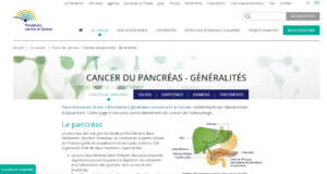
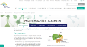
The pancreas is an organ that forms part of the digestive system. Pancreatic cancer is an uncontrolled growth of malignant tumorous cells within the pancreas.
Since there are different cell types present within the pancreas, we distinguish two basic types of pancreatic cancer.
Adenocarcinoma is the most prevalent form of pancreatic cancer. It originates in the exocrine glands.
Less frequently occurring types of pancreatic cancer are:
In Belgium, around 1,500 patients per year are diagnosed with pancreatic cancer. Although a lot of resources are poured into research towards treatment, pancreatic cancer remains one of the least curable cancers. One of the main challenges with pancreatic cancer is the fact that the disease often does not get diagnosed until it is at a relatively advanced stage and has already metastasised.
Most patients with pancreatic cancer do not notice any symptoms during the early stages of the disease. The cancer only becomes apparent at a late stage, which means that treatment also starts relatively late, thus greatly reducing survivability.
The first things that patients notice are:
Further symptoms include:
Despite many years of intensive research, scientists have yet to pinpoint the exact cause of pancreatic cancer. A few risk factors are:
When a doctor suspects a patient may have developed pancreatic cancer, he or she will be referred to an oncologist. The oncologist will then conduct investigations such as blood tests, echo tests and CT scans to confirm the diagnosis.
As soon as the diagnosis has been confirmed, further investigations are needed in order to establish how far the pancreatic cancer has advanced, what type it is and whether the disease has metastasised or not. This is done by MRI or MRCP scans, endo-echography and possible laparoscopy or an ERCP. Subsequently, a treatment strategy is decided upon.
An ERCP, endoscopic retrograde cholangio-pancreaticography, is an examination of the biliary and pancreatic tract. This is done by means of an endoscope, a flexible hose with a micro-camera attached to it. An ERCP can provide results that may not be obtained by traditional imaging technology. Usually, an ERCP investigation is only conducted by specialists when it is anticipated that a medical procedure has to take place during the examination.
Pinpointing the exact stage of the pancreatic cancer is invaluable when it comes to establishing a prognoses and deciding on a treatment plan.
Five stages
The differentiation of the tumour is an important factor in establishing a prognosis and treatment. This can be determined on the basis of a biopsy. A biopsy involves the removal of a small bit of tissue, which can be examined under a microscope. Differentiation determines the degree of mutation in the cancerous cells.
Due to the fact that pancreatic cancer almost always gets diagnosed at a late stage, only 25% of all patients will receive curative treatment. But in most cases, treatment is limited to palliative care. In Belgium, patients with metastasised pancreatic cancer that has reached lymph nodes outside the operable area are not deemed suitable for operation. The same principle applies to patients where imaging has shown the tumour to be inoperable.
If examinations show that the tumour can be removed, then the patient is operated on and receives subsequent chemotherapy and/or radiotherapy to reduce the risk of recurrence or metastasis. The latter two treatments also form part of palliative care. Furthermore, if the patient’s cancer expresses a BRCA mutation, then PARP inhibitors can be used. Research into targeted therapy is currently ongoing.
Currently, there are no dedicated patient groups for pancreatic cancer in Belgium. Flemish patients are referred to the Dutch organisation, whereas French speakers can find information with the French patient association AFRCP.